Novel Light‑Based Imaging Reveals Hidden Activity in Blood Immune Cells
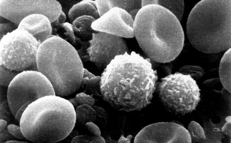
Researchers have introduced an innovative optical imaging approach capable of uncovering previously invisible functional signals within peripheral blood mononuclear cells (PBMCs), the varied immune cells routinely obtained from a routine blood draw. Detailed in a recent pre‑print, the method promises deeper insight into how these cells react to infection, autoimmunity and cancer without requiring invasive techniques.
PBMCs—comprising lymphocytes and monocytes—are a mainstay of immunology studies because they provide a real‑time snapshot of the body's immune landscape. Historically, scientists have depended on flow cytometry, gene‑expression profiling or cytokine assays to infer cellular activity. Although powerful, these strategies often need labeling, bulk analysis or extensive processing that can obscure dynamic, single‑cell behavior.
The newly described platform leverages sophisticated light‑scattering and fluorescence‑free microscopy to obtain high‑resolution, live‑cell images of PBMCs in real time. By scrutinizing minute changes in optical density and intracellular motion, the system can tell resting cells from activated ones even when traditional markers remain unchanged. In pilot tests, the imaging setup revealed distinct activity patterns in cells from individuals with viral infections compared with healthy donors.
The authors highlight several practical benefits. Because no fluorescent tags are required, sample preparation is quicker and the risk of staining‑induced artifacts disappears. The label‑free nature also permits repeated measurements on the same sample, paving the way for longitudinal tracking of disease progression or therapeutic response.
Beyond basic research, clinicians may eventually employ the technique to spot early immune dysregulation in autoimmune diseases or to assess the efficacy of cancer immunotherapies. Detecting subtle immune shifts before overt symptoms emerge could enable interventions at an earlier stage, aligning with the growing push toward personalized medicine.
Nonetheless, the approach is still undergoing validation. Larger cohorts are necessary to verify that the optical signatures reliably correspond to specific disease states across diverse populations. Incorporating the method into current diagnostic pipelines will also demand standardized imaging protocols and robust software for data analysis.
If forthcoming trials substantiate its value, the technology could become a routine element of blood‑based diagnostics, complementing existing assays and providing a non‑invasive glimpse into the immune system’s concealed activity. The research community will be watching closely as the concept advances from proof‑of‑concept toward possible clinical use.



Comments (0)
Be the first to comment.
Join the discussion